
How Is Lung Cancer Diagnosed? 10 Powerful Diagnostic Facts
Understanding how is lung cancer diagnosed can help people know what to expect when symptoms or an abnormal scan raise concerns. Lung cancer diagnosis usually involves several steps rather than one single test. Doctors may begin with a medical history and physical examination, followed by imaging tests, laboratory investigations, and procedures that collect tissue for examination.
For people undergoing evaluation in Thrissur, the diagnostic process may vary depending on symptoms, overall health, imaging findings, and individual risk factors. The main goal is to determine whether cancer is present, identify the specific type of lung cancer, and establish how far it may have spread.
A clear diagnosis is important because many lung conditions can cause similar symptoms. Infections, inflammation, tuberculosis, and other respiratory diseases can sometimes produce findings that resemble lung cancer. Therefore, proper testing is necessary before a diagnosis is confirmed.
How Is Lung Cancer Diagnosed Initially
When discussing how is lung cancer diagnosed, the process usually begins when a person develops suspicious symptoms or when an abnormality is discovered during an imaging test performed for another reason.
Possible warning signs may include:
- A persistent or worsening cough
- Coughing up blood
- Chest pain
- Shortness of breath
- Unexplained weight loss
- Persistent tiredness
- Recurrent chest infections
- Hoarseness
- Reduced appetite
- Wheezing
These symptoms do not automatically mean that a person has lung cancer. Many non-cancerous conditions can produce similar problems.
Medical History
The healthcare professional may ask about the person’s symptoms, how long they have been present, smoking history, occupational exposures, previous lung conditions, family medical history, and general health.
Information about exposure to substances such as asbestos, radon, silica, or certain workplace chemicals may also be relevant.
Physical Examination
A physical examination may include listening to the lungs, checking breathing patterns, examining lymph nodes, and looking for signs that could indicate respiratory disease.
If further investigation is necessary, imaging tests are generally the next step.
Chest X-Ray in Lung Cancer Diagnosis
A chest X-ray is one of the initial imaging tests that may be performed when lung-related symptoms are present.
It can sometimes identify:
- An abnormal lung mass
- Areas of collapsed lung
- Fluid around the lungs
- Unusual shadows
- Changes in lung structure
However, a chest X-ray alone cannot confirm lung cancer.
Small tumors may not always be visible, and some abnormalities seen on an X-ray may be caused by infections, scar tissue, or other non-cancerous conditions.
If the X-ray reveals something suspicious, a CT scan may be recommended for a more detailed examination.
How Is Lung Cancer Diagnosed With a CT Scan
A computed tomography or CT scan plays an important role when explaining how is lung cancer diagnosed.
A CT scan uses X-rays and computer processing to create detailed cross-sectional images of the chest. It provides considerably more information than a standard chest X-ray.
CT imaging may help identify:
- The size of a lung nodule or tumor
- The exact location of an abnormality
- Enlarged lymph nodes
- Multiple lung nodules
- Involvement of nearby structures
- Possible spread within the chest
Why Is a CT Scan Important?
A CT scan can detect smaller abnormalities that may not appear clearly on a conventional chest X-ray.
However, imaging cannot always determine whether an abnormal growth is cancerous. A suspicious nodule may still require additional testing, particularly a biopsy.
PET-CT Scan for Evaluating Lung Cancer
A positron emission tomography-computed tomography, or PET-CT, scan combines metabolic information from a PET scan with detailed anatomical information from a CT scan.
A small amount of radioactive tracer is administered before the scan. Areas with increased metabolic activity can absorb more of the tracer and appear more prominently.
Because many cancer cells have increased metabolic activity, PET-CT imaging can help evaluate suspicious areas.
What Can a PET-CT Scan Show?
It may help doctors:
- Assess a suspicious lung mass
- Evaluate lymph nodes
- Look for possible cancer spread
- Determine the extent of disease
- Select appropriate areas for biopsy
- Support treatment planning
PET-CT findings alone do not necessarily confirm cancer. Infection and inflammation can also produce increased metabolic activity.
Biopsy: An Important Step in Confirming Lung Cancer
A biopsy is generally necessary to confirm whether suspicious lung tissue contains cancer cells.
During a biopsy, a small sample of cells or tissue is collected. A pathologist then examines the sample under a microscope.
When people ask how is lung cancer diagnosed accurately, tissue examination is one of the most important parts of the process.
Several biopsy techniques may be used depending on where the suspicious area is located.
Needle Biopsy
A needle biopsy may be recommended when a suspicious tumor is located toward the outer part of the lung.
Imaging guidance, such as CT, can be used to guide a thin needle through the chest wall and into the abnormal area.
A tissue sample is then removed for laboratory examination.
Lymph Node Biopsy
If enlarged or suspicious lymph nodes are present, tissue may be collected from them to determine whether cancer cells have spread beyond the primary lung tumor.
Bronchoscopy for Lung Cancer Diagnosis
Bronchoscopy is a procedure used to examine the airways inside the lungs.
A thin, flexible instrument called a bronchoscope is usually passed through the nose or mouth and into the airways.
The bronchoscope contains a light and camera, allowing the airways to be viewed directly.
What Happens During Bronchoscopy?
If an abnormal area is identified, small instruments can be passed through the bronchoscope to collect tissue or cell samples.
Bronchoscopy may be particularly useful for tumors located near the larger central airways.
Samples collected during the procedure are sent to a laboratory for examination.
Endobronchial Ultrasound
Endobronchial ultrasound, commonly called EBUS, combines bronchoscopy with ultrasound imaging.
It can help visualize lymph nodes and structures near the airways.
A needle can then be guided into selected lymph nodes to collect samples. This information can help both diagnose lung cancer and determine whether it has spread to nearby lymph nodes.
Laboratory Testing of Lung Tissue
Once a biopsy sample has been collected, laboratory analysis becomes an important part of determining how is lung cancer diagnosed and classified.
A pathologist examines the cells to determine whether they are cancerous.
If cancer is identified, the laboratory may classify it into one of the major lung cancer categories.
Non-Small Cell Lung Cancer
Non-small cell lung cancer, or NSCLC, is the most common broad category.
Major subtypes include:
- Adenocarcinoma
- Squamous cell carcinoma
- Large cell carcinoma
Small Cell Lung Cancer
Small cell lung cancer, or SCLC, generally behaves differently from non-small cell lung cancer and can grow and spread relatively quickly.
Accurately identifying the type is essential because treatment strategies differ between different forms of lung cancer.
Molecular and Biomarker Testing
Modern lung cancer diagnosis may extend beyond simply identifying cancer cells.
For some patients, particularly those with certain types of non-small cell lung cancer, tumor tissue may undergo molecular or biomarker testing.
These tests look for specific genetic or molecular characteristics within the cancer.
Depending on the clinical situation, testing may evaluate alterations involving genes such as:
- EGFR
- ALK
- ROS1
- BRAF
- KRAS
- MET
- RET
Other biomarkers may also be evaluated depending on the cancer type and available testing.
PD-L1 Testing
PD-L1 testing may be performed in some cases to help determine whether certain immunotherapy treatments could be appropriate.
Biomarker results can therefore provide valuable information for treatment planning after the diagnosis has been established.
Blood Tests During Lung Cancer Evaluation
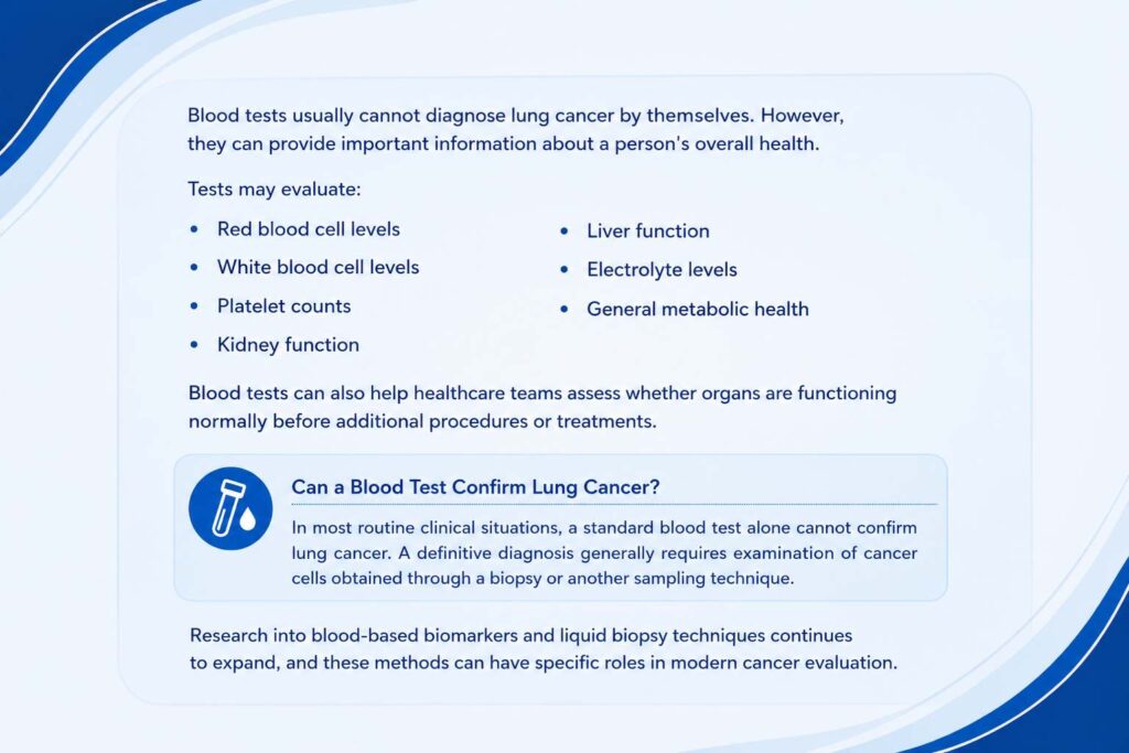
Blood tests usually cannot diagnose lung cancer by themselves. However, they can provide important information about a person’s overall health.
Tests may evaluate:
- Red blood cell levels
- White blood cell levels
- Platelet counts
- Kidney function
- Liver function
- Electrolyte levels
- General metabolic health
Blood tests can also help healthcare teams assess whether organs are functioning normally before additional procedures or treatments.
Can a Blood Test Confirm Lung Cancer?
In most routine clinical situations, a standard blood test alone cannot confirm lung cancer.
A definitive diagnosis generally requires examination of cancer cells obtained through a biopsy or another sampling technique.
Research into blood-based biomarkers and liquid biopsy techniques continues to expand, and these methods can have specific roles in modern cancer evaluation.
ests Used to Determine Whether Lung Cancer Has Spread
Once cancer has been confirmed, additional testing may be needed to establish its stage.
Staging describes the extent of cancer in the body.
Tests used during staging may include:
- CT scans
- PET-CT scans
- MRI scans
- Bone imaging in selected cases
- Lymph node sampling
- Additional biopsies
Brain MRI
Lung cancer can sometimes spread to the brain. An MRI may therefore be recommended in certain patients to look for abnormalities that could indicate metastatic disease.
Why Staging Matters
Staging helps determine:
- How large the tumor is
- Whether nearby lymph nodes are involved
- Whether cancer has reached nearby tissues
- Whether it has spread to distant organs
This information is essential when deciding which treatment approaches may be appropriate.
Common Causes and Risk Factors for Lung Cancer
Although understanding how is lung cancer diagnosed is the primary concern during evaluation, doctors also consider possible risk factors as part of the medical history.
Smoking is a major risk factor for lung cancer, but people who have never smoked can also develop the disease.
Other recognized risk factors may include:
- Secondhand tobacco smoke
- Radon exposure
- Asbestos exposure
- Occupational carcinogens
- Air pollution
- Previous radiation exposure
- Family history
- Increasing age
Having one or more risk factors does not mean that lung cancer will definitely develop.
Similarly, the absence of obvious risk factors does not completely exclude the possibility.
Types of Lung Cancer Identified During Diagnosis
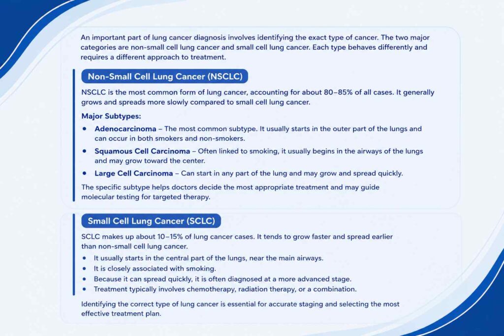
An important part of understanding how is lung cancer diagnosed involves knowing why doctors need to identify the exact cancer type.
The two major categories are non-small cell lung cancer and small cell lung cancer.
Non-Small Cell Lung Cancer
NSCLC represents the majority of lung cancers. Adenocarcinoma and squamous cell carcinoma are among its common subtypes.
Treatment depends on factors including stage, tumor characteristics, molecular findings, and overall health.
Small Cell Lung Cancer
Small cell lung cancer is less common but often grows and spreads more rapidly.
Because its biological behavior differs from NSCLC, its staging and treatment approach may also differ.
Symptoms That May Lead to Lung Cancer Testing
Lung cancer may not cause noticeable symptoms during its earliest stages. Symptoms can become more apparent as the disease develops.
Potential symptoms include:
- Persistent coughing
- Changes in an existing cough
- Blood in sputum
- Chest discomfort
- Difficulty breathing
- Unexplained weight loss
- Fatigue
- Reduced appetite
- Hoarseness
- Repeated respiratory infections
These symptoms have many possible causes. Persistent or unexplained symptoms should be medically evaluated rather than being assumed to represent cancer.
Why Early Lung Cancer Diagnosis Matters
Knowing how is lung cancer diagnosed also highlights the importance of timely evaluation.
Finding lung cancer at an earlier stage may increase the range of treatment options available.
Potential benefits of timely diagnosis include:
- Earlier identification of suspicious abnormalities
- More accurate staging
- Faster treatment planning
- Greater opportunity for potentially curative treatment in eligible cases
- Identification of relevant molecular biomarkers
- Better understanding of the disease
Early diagnosis does not guarantee a particular outcome, but it can significantly influence treatment options.
What Happens After Lung Cancer Is Diagnosed
After laboratory testing confirms cancer, doctors combine information from pathology, imaging, staging investigations, and overall health assessments.
The treatment plan depends on several factors.
These include:
- Lung cancer type
- Cancer stage
- Tumor location
- Molecular characteristics
- Overall health
- Lung function
- Other medical conditions
- Individual treatment considerations
Treatment Options After Lung Cancer Diagnosis
Treatment is not part of the diagnostic process itself, but diagnosis provides the information needed to select an appropriate approach.
Depending on the type and stage of lung cancer, treatment may involve:
Surgery
Surgery may be considered for some localized non-small cell lung cancers when the tumor can be removed and the patient is medically suitable.
Radiation Therapy
Radiation therapy uses high-energy radiation to damage cancer cells. It may be used alone or in combination with other treatments.
Chemotherapy
Chemotherapy uses medicines that destroy or interfere with rapidly dividing cancer cells.
Targeted Therapy
Targeted treatments act against particular molecular abnormalities found in some cancer cells. Molecular testing helps determine whether certain targeted therapies may be relevant.
Immunotherapy
Immunotherapy helps the immune system recognize or attack cancer cells. Biomarker testing and other clinical factors can influence whether immunotherapy is considered.
Treatment decisions are individualized and depend on the complete diagnostic and staging results.
Tips for Preparing for Lung Cancer Diagnostic Tests
The diagnostic process can involve several appointments and different tests. Being organized can make it easier to understand what is happening.
Helpful steps include:
- Keep a list of symptoms and when they began.
- Bring information about previous medical conditions.
- Provide an accurate smoking and exposure history.
- Carry previous imaging reports when available.
- Ask what each test is designed to determine.
- Ask whether preparation is required before a procedure.
- Keep copies of important reports.
- Write down questions before appointments.
- Ask when test results are expected.
Understanding the purpose of each investigation can make the diagnostic process easier to follow.
Conclusion
Understanding how is lung cancer diagnosed means recognizing that diagnosis is usually a multistep process. Imaging tests such as chest X-rays, CT scans, PET-CT scans, and MRI can identify suspicious abnormalities and evaluate their extent, while bronchoscopy, needle biopsy, or lymph node sampling can provide tissue for laboratory examination.
Pathology confirms whether cancer is present and determines its type. Molecular testing may then provide additional information that helps guide treatment. Staging tests establish how far the disease has spread and support appropriate treatment planning.
Anyone experiencing persistent respiratory symptoms or undergoing investigation for an abnormal lung finding should follow the recommended diagnostic evaluation. Access to appropriate imaging, biopsy, pathology, and staging services in Thrissur can support an accurate understanding of the condition and help guide the next stage of care. According to the American Cancer Society – Tests for Lung Cancer, imaging tests, biopsies, and laboratory examinations are commonly used to diagnose lung cancer and determine its extent.
Frequently Asked Questions

Dr. Bibin Francis
Dr. Bibin Francis is a dedicated medical oncologist with expertise in chemotherapy, immunotherapy, targeted therapy, and personalized cancer treatment planning for a wide range of malignancies.
Jubilee Mission Medical College & Research Institute, Thrissur, Kerala